Failure is Not an Option
The image above has, like many photos, a story behind it. Often non-photographers look at photos and do not understand what went into the making of a good photo. When working with the microscope, things are even more alien as there is a bunch of additional considerations that need to be taken into account before you can even begin turning on the camera.
Lets start at the beginning. Not all good photographs can be had at first attempt. Sometimes you have to fail (many times) before you can succeed. Here is the best I could do 4 years ago:
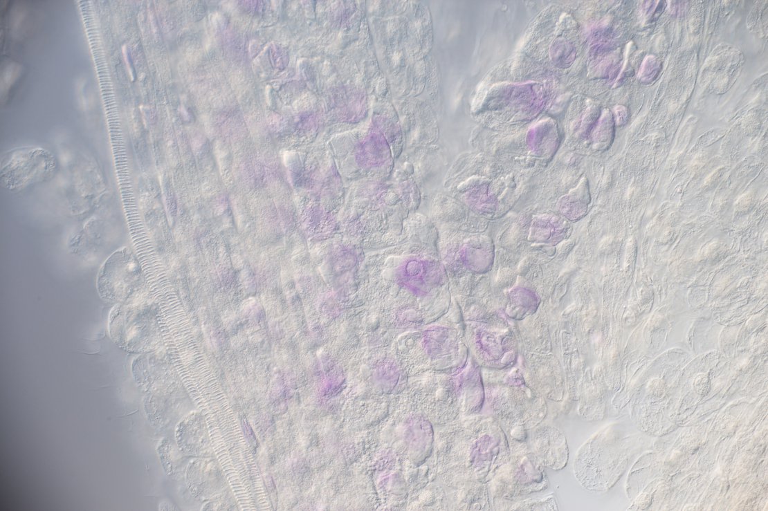
To understand the failure here, I need to give a quick overview of DNA staining. In order to observe mitosis (the process of cell division and replication), one needs to stain the DNA of a cell using special chemicals in order for the chromosomes to be visible under the light microscope. Once stained, the root tip is squashed under the microscope slide to spread out the layers of cells in order to view individual cells clearly. Many things can go wrong. In the image above, one can hardly see any nuclei - they have been stained poorly. So I first had to correct my approach. Since this is not an article about staining techniques, I'll skip to the improved result:
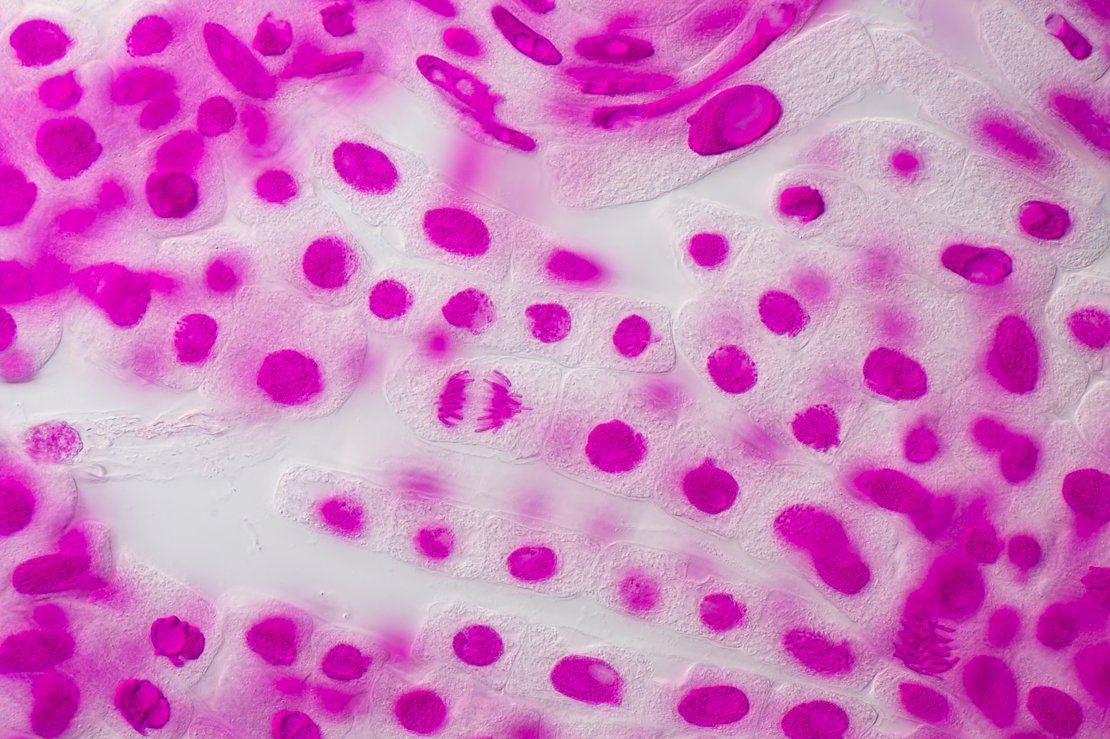
Clearly, this is a marked improvement. The staining is good, the root tip is in good condition and mitosis is clearly present. However, there are a couple of issues:
- The cells are not in a single layer, making the overall impact of the image less dramatic.
- The staining is a bit too intense, causing some detail to be lost in a sea of magenta
- I am not convinced DIC is the correct illumination to show mitosis. It does show the cells much better, but it adds visual distractions.
So 4 years later I decided to give it another go. My goals were to improve on all the above-mentioned issues, as well as getting closer to see more detail. Here is my first second attempt:
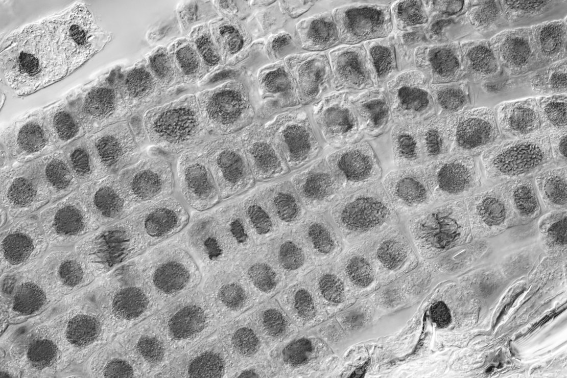
Not what you want to see when you are supposed to be 4 years wiser! The image is in black & white as that was the only way I thought of at the time, to improve on contrast. It is clear that my root tip is in a good condition and showing active mitosis, however somewhere down the line my technique was flawed, resulting in an image that is vastly inferior to my first attempt 4 years ago.
I spent some time improving my staining technique, squashing of the root tip as well as image processing. Here is what I managed to get after 2 days:
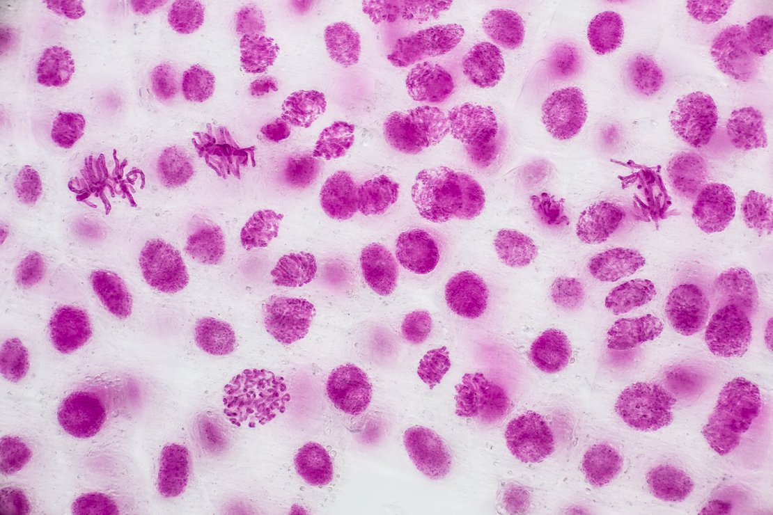
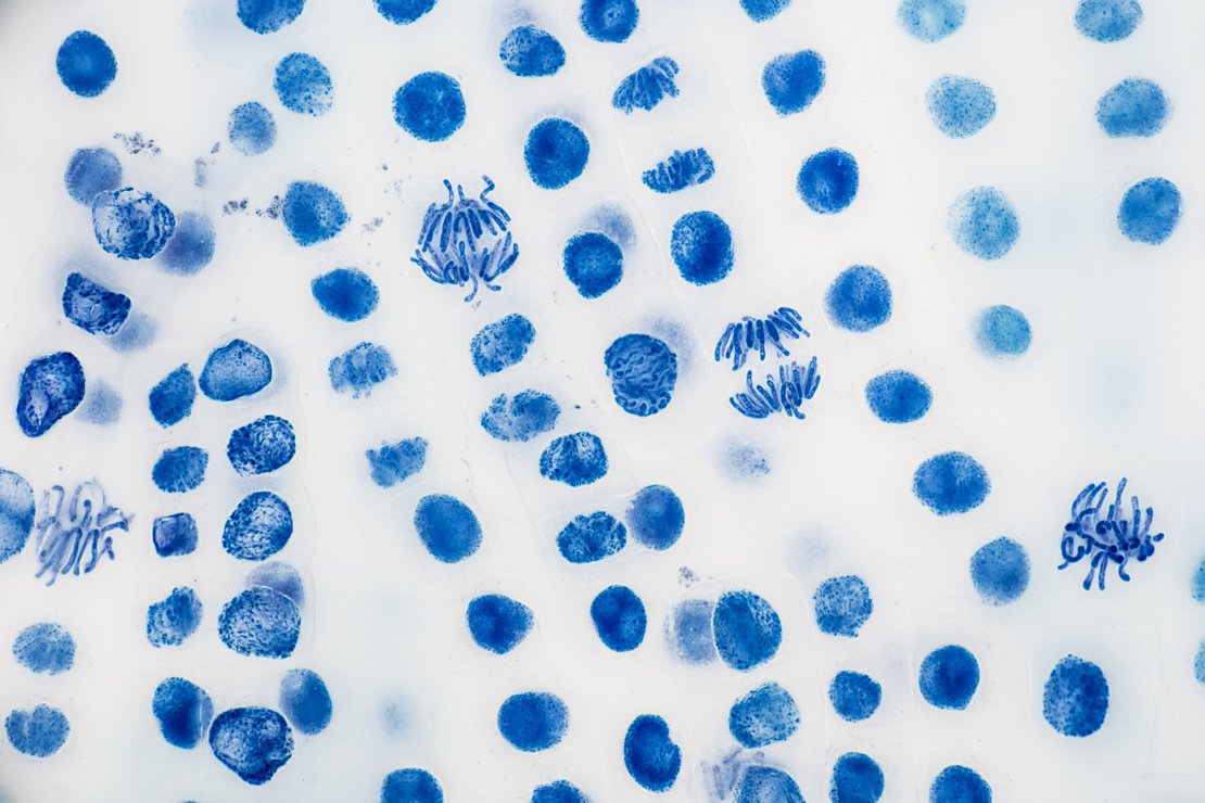
This is much better, even than my first attempt. The individual chromosomes are clearly visible, the double layer of cells are not too distracting as they are part of the bokeh, and the magenta is not overpowering and wiping out the detail in the image. But I knew I could do even better. I decided to switch to a different chemical stain, worked a bit on my preparation and processing techniques and the image I have started with was the result:
There are no distracting cells in the background, the chromosomes in the cells undergoing mitosis are clear and well defined, and a bit more detail can be seen since I was using a 100x NA1.4 objective with oil - basically the highest resolution a light microscope can achieve (excluding fluorescence).